INTRODUCTION
HSV is an envelop, DNA-comtaining virus morphologically similar to the other members of the genus Herpesviridae.Two antigenically distinct types are recognized, designated type 1 and type 2.
HSV type 1 and 2 are frequently implicated in superficial infections of the oral cavity, the skin, the eye and the genitalia, Infections of the central nervous system (meningoencephalitis)and severe generalized infection in the neonate of immunocompromised patient are also seen, though more rarely. After the primary infection been resolved, the virus may exist in a latent form in nervous tissue, from where it may re-emerge, under certain conditions, to cause a recurrence of the symptoms.
The classical clinical presentation of genital herpes starts with widespread multiple painful macules and papules, which then mature into clusters of clear, fluid-filled vesicles and pustules. The vesicles rupture and form ulcers. Skin ulcers crust, whereas lesions on mucous membranes heal without crusting. In women, the ulcers occur at the introitus, labia, perineum, or perianal area. Men usually develop lesions on the penial shaft or glans. The patient usually develops tender inguinal adenopathy. Perianal infections are also common in MSM. Pharyngitis may develop with oral exposure.
Serology studies suggest that 50 million people in the United States have genital HSV infection. In Europe, HSV-2 is found in 8-15% of the general population. In Africa, the prevalence rates are 40-50% in 20-year-olds. HSV is the leading cause of genital ulcers. HSV-2 infections at least doubles the risk of sexual acquisition of human immunodeficiency virus (HIV) and also increases transmission.
Until recently, viral isolation in cell culture and determination of the type of HSV with fluorescent staining has been the mainstay of herpes testing in patients presenting with characteristic genital lesions. Besides PCR assay for HSV DNA has been shown to be more sensitive than viral culture and has a specificity that exceeds 99.9%. But these methods in clinical practice are currently limited, because the cost of the test and the requirement for experienced, trained technical staff to perform the testing restrict their use.
There are also commercially available blood tests used for detecting Type Specific HSV antibodies, but these serological testing cannot detect primary infection so they can be used only to rule out recurrent infections. This novel antigen test can differentiate other genital ulcer diseases with genital herpes, such as syphilis and chancroid, to help the early diagnosis and therapy of HSV infection.
PRINCIPLE
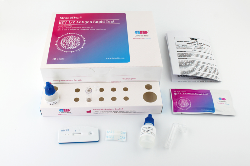
The HSV antigen Rapid Test Device has been designed to detect HSV antigen through visual interpretation of color development in the internal strip. The membrane was immobilized with anti Herpes simplex virus monoclonal antibody on
the test region. During the test, the specimen is allowed to react with colored monoclonal anti-HSV antibody colored particals conjugates, which were precoated on the sample pad of the test. The mixture then moves on the membrane by capillary
action, and interacts with reagents on the membrane. If there were enough HSV antigens in specimens, a colored band will form at the test region of the membrane. Presence of this colored band indicates a positive result, while its absence indicates
a negative result. Appearance of a colored band at the control region serves as a procedural control. This indicates that proper volume of specimen has been added and membrane wicking has occurred.

